Research — General Overview
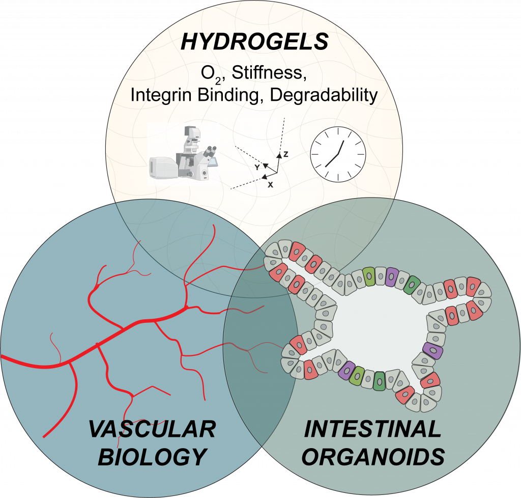
We use spatiotemporally tunable hydrogels to culture cells and study how evolving microenvironmental properties regulate tissue and organ growth. We use these studies to understand elements of fundamental biology, but also to identify characteristics of injured or diseased microenvironments that can be targeted to treat disease.
Our primary focus is on studying gut organoids, with other projects involving vascular cells. Our methods can be modified for culture or co-culture of other cell and organoid types to continually add complexity into our models or learn more about the biology and pathobiology of other types of tissues.

Designer Cell Niches
We learn from nature to build better tissue specific niches.
We take what is known about tissue-specific microenvironments and build our own, highly controllable versions in the lab. We’re interested in how characteristics of tissue-specific microenvironments, including matrix composition and mechanics, impact morphogenesis. To date, we have focused on building “healthy” tissue niches to understand some fundamentals of intestinal biology. We have ongoing work designing intestinal developmental niches, and plans to create disease-relevant microenvironments.
Selected Papers, Conference Proceedings, and Preprints:
Hushka EA, Blatchley MR, Macdougall LJ, Yavitt FM, Kirkpatrick BE, Bera K, Dempsey PJ, Anseth KS. Fully synthetic hydrogels promote robust crypt formation in intestinal organoids. bioRxiv. (link)
Blatchley MR, Hall F, Wang S, Pruitt HC, Gerecht S. Hypoxia and matrix viscoelasticity sequentially regulate endothelial progenitor cluster-based vasculogenesis, Science Advances, 5 (3), eaau7518, 2019. (link)
Spatiotemporal Tissue Engineering
We use light to guide tissue morphogenesis and construct tissues with user-controlled geometries.
Microenvironmental dynamics are crucial in homeostasis, regeneration, development, and disease. In homeostasis, cells do their thing to keep tissues functioning. This can include remodeling their environment and changing their identity when needed. These functions change in an elegant way throughout development, resulting in more mature and functional tissues. In response to injury, and when things go wrong in disease, the niche dynamics shift in beneficial ways for regeneration or detrimental ways in disease. We recreate these positive and negative shifts to understand how cells respond.
Papers, Conference Proceedings, and Preprints:
Blatchley MR, Bera K, Kirkpatrick BE, Yavitt FM, McGrath PS, Dempsey PJ, Anseth KA. Spatiotemporally controlled matrix softening facilitates determinisitic crypt formation in iPSC-derived intestinal organoids. In preparation.
Yavitt FM, Kirkpatrick BE, Blatchley MR, Speckl KF, Mohagheghian E, Moldovan R, Wang N, Dempsey PJ, Anseth KS. In situ modulation of intestinal organoid epithelial curvature through photoinduced viscoelasticity directs crypt morphogenesis. Science Advances, 9 (3), eadd5668, 2023. (link)
Advanced Imaging & Characterization Techniques
We take lots of pictures of organoids and biomaterials to understand how they change over time and how cells interact with and remodel their surroundings.
Imaging provides spatial context, which is crucial in understanding oragnoid biology. We use a variety of imaging techniques to characterize organoids and material properties.
Papers, Conference Proceedings, and Preprints:
Blatchley MR*, Günay KA*, Yavitt FM, Hawat EM, Dempsey PJ, Anseth KS. In situ super-resolution imaging of organoids and extracellular matrix interactions via photo-transfer by allyl sulfide exchange expansion microscopy (PhASE-ExM), Advanced Materials, 2109252, 2022. (link)
Blatchley MR, Günay KA, Yavitt FM, Dempsey PJ, Anseth KS. Synthetic hydrogels to study ECM dynamics of intestinal organoids, Gastroenterology, 160(6), S434-S434. (link)
Please reach out if you are interested in collaborating and check our OPPORTUNITIES page for open positions in the lab.